Wednesday, March 2, 2011

EKG Lab, Cardiology Lab, Heart Disection, Etc.

Andrew


Over the past couple weeks we have been learning about the cardiovascular system in numerous labs. We have learned the valves of the heart and the basic parts and even the blood flow of the heart. In another Lab we study each of our group members heart pulse and blood pressure. To do this you can use older techniques or newer but we went newer because we could never be a doctor with a stethoscope. We used some new technique that did all the work for us and graphed it onto a graph on the computer which we found that we were all relatively the same and pretty healthy. Now the the graph at the left are from the EKG Lab which is where we did a time of each of our heart beat over the time of five seconds with some wires hooked up on our wrist and and forearm to calculate the pulse. The ones with our names are are basic with it normal and found that mine and Danial heart are nearly identical and Andrew's made and big jump and fell to a stable normal level. Then the name and Reversed is when we switched the red and green wire to look at it a different way. Which makes it seem your heart beat is out of whack but it's no big deal and the machines is the reason for all the great heart data. 
Now the heart dissection is where we cut a cow, sheep, and pig heart by splitting in half to see the inside of the ventricles and atriums and took the measurements of every basic part of the heart the valves and the size of the ventricles and atriums. Our data showed the pig and cow heart had the biggest walls, valves, just basic parts of the heart were relatively big, to me this help us to learn and study the heart to prepare us for our quiz we ended up having. To see this data the graph of this lab is at the bottom of the page.
Overall I believe that all of the study of the heart help to teach and understand a little more about the flow of blood and what certain parts of the heart does and how the arteries and veins play an huge part in the system as arteries carry blood from the heart and veins carry blood to the heart. Also, the pig picture of understand the process of how the objective of life is carried out with these processes of the cardiovascular system. Overall a great study of the heart took place and I believe it benefited me greatly as, well as the class.


Andrew Reversed
Daniel 


 Daniel Reveresed


Brian


Brian Revered


Cow Heart


Monday, January 24, 2011

Leech Neurophysiology Lab

Purpose: The purpose of this lab is to identify the different types of cells located in the leech.

Hypothesis: If you probe correctly then you will be successful in finding the glands and then the resources will help you identify them.

Materials:
Feather
Probe
Forceps
Scissors
Pins
Scalpel
Dissection Tray
Leech Tank
20% Ethanol
Leech Tongs
Dissection Microscope
Micromanipulator
Oscilloscope
Leech
Procedure: The procedure of this lab you anesthetize( so it will not move around) the leech and prepare it the dissection, by pinning it down on the tray. Then begin to cut and remove the guts from the leech, after you cut out the ganglion window and isolate one to study. After that you cut out the ganglion sinus under a microscope and probe and begin to identify ganglion sensory glands. That is the procedure taking in this lab of what you need to do to succeed.

Results- My results showed how to identify glands correctly by the help of the machine and atlas which help to identify them correctly too. My hypothesis was correct in a way because you will find the glands if properly probe and use the resources correctly, but the different utensils played a role in fighting some.

Conclusions- In this lab I identify and found every gland correctly thanks to the resources and that is the reason I was correct, to me the only thing is that the lab is crazy how using the different objects, to find out the different glands by using the probe, feather, and forceps. That way the UV will detect a cell and then find one.

Friday, December 17, 2010

Skeletal Muscle Tissue

This is the skeletal muscle tissue shown in the photo at the right and this skeletal muscle plays an important role in the human body and I will discuss these functions.
1. First I will discuss the first the functions as a whole, one it helps to produce movement which is the muscle contraction and help you to respond quickly.
2. Also, they help to maintain posture and this is because they make tiny adjustments which help us maintain our body posture or seated up.
3. These muscles help to maintain the stabilization of joints because the muscles pulling on bones causes movements  and helps to strengthen and stabilize them.
4. Last it helps to generate heat because they contract which helps to maintain normal body temperature and it's the most important muscle type for generating the heat.
Now just for some individual structures of what they are:
Muscle- it consists of hundreds to thousands of muscle cells, plus connective tissue wrappings, blood vessels, and nerve fibers
Fascicle- is a portion of the muscle and is a discrete bundle of muscle cells, segregated from the rest of the muscle by a connective tissue sheath
Muscle fiber- is a elongated multinucleate cell; which has a banded appearance.
Myofibril or fibril- is a rodlike contracile element; they occupy most of the muscle cell volume; bands of adjacent myofibrils are aligned; and composed of sarcomeres arranged end to end.
Sarcomere- it's the contractile unit, composed of myofilaments made up of contractile proteins
Myofilament or filament- there are two types thick and thin the thick contain bundled mysoin molecules; and thin contain actin molecules.

This is an important part of the human body to me because of those top four functions: produce movement, maintain posture, stabilize joints, and help to maintain temperature. To me I would have to say it's extremely important without this there would be no life for us because not having movement would be the worst filling in the world and that's why to me.

EKG Lab

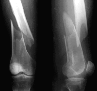
Types of Fractures

Comminuted Fracture- Bone fragments into three or more pieces, particularly common in the aged, whose bones are more brittle.


Compression Fracture- Bone is crushed, is common in porous bones (i.e., osteoporotic bones) subjected to extreme trauma, as in a fall.


Spiral Fracture- Bagged break occurs when excessive twisting forces are applied to a bone, is common sports fracture.


Epiphyseal Fracture- Epiphysis separates from the diaphysis along the epiphyseal plate, and tends to occur where cartilage cells are dying and calcification of the matrix is occurring.


  Depressed Fracture- Broken bone portion is pressed inward, typical of skull fractures.


Greenstick Fracture- Bone breaks incompletely, much in the way a green twig breaks. Only one side of the shaft breaks; the other side bends, is common in children, whose bones have relatively more organic matrix and are more flexible than those of adults.


Now we will discuss some more about these fractures and little of bone repair. It's important to know the type of fracture you have because that why the doctor can give you the proper treatment and that why the bone will heal properly. So that is why it's important to understand and know what type of fracture it's that you have. Now to discuss some treatment, to treat a fracture you must be by reduction or the realignment of the broken bones. There are two types of reduction closed and open, closed is when the bone ends are put into position by the doctor's hands, and in open the bone ends are secured by pins or wires. Then of course everyone knows then it's immobilized by a cast or traction. Then it takes six to eight weeks to heal and that is important to know that way you know how your bones heal and understand what goes into the healing process.

Directional Body Terms

Superior (cranial) Toward the head or end or upper part of a structure or the body; above
Inferior (caudal) Away from the head end or toward the lower part of  a structure or the body; below
Anterior (ventral) Toward or at the front of the body; in front of
Posterior (dorsal) Toward or at the back of the body; behind
Medial- Toward or at the midline of the body; on the inner side of
 Proximal- Closer to the origin of the body part or the point of attachment of a limb to the body trunk
Distal- Farther from the origin of a body part or the point of attachment of a limb to the body trunk
Superficial (external) Toward or at the body surface
Deep (internal) Away from the body surface; more internal
Intermediate- Between a more medial and a more lateral structure
Lateral- Away from the midline of the body; on the outer side of


Why are the Directional Body Terms important to science, I believe this is because it helps to direct where the body parts are on the body and would help to discuss to others doctors which way to cut or where something is located. Another reason why I believe these terms are helpful is just to know the language of Anatomy which I think plays a role of how and why doctors learn how to discuss this. That to me is why these terms even play any role at all in the world and that's why I think it's important in the world of Anatomy.

Friday, October 15, 2010

Organization of the body

This is the process of what builds up to an organism and the final step is the organismic level which is all structural promotions working to maintain life.
1. Chemical Level
    a. atoms (tiny blocks of matter) combine to form molecules (water, protein, etc.) they in turn form organelles ( basic stuff of microscopic cells)

2. Cellular Level
     a. They are made up of molecules and enhance to create tissues.

3. Tissue Level (combination similar cells)
    a. groups of small cells have common fuction
         1. Types
              a. Epitheium ( covers bodies surface and lines cavities)
              b. Muscle ( movement)
              c. Connective Tissue ( supports protects organs)
              d. Nervous Tissue ( rapid communication by electrical impulses)

4. Organ Level ( different types of tissue)
   a. at least two types of tissue and complex functions are possible
   b. have specialized functions
                                    
                                                     5. Organ System Level ( work together for common purpose)
                                                        Ex: Respiratory, Muscular, Nervous, or Digestive systems
                                                    
                                                      6. Organismal Level
                                                       a. all structural promotions working to maintain life
The organization of the body is very important because it builds up from atoms all the way to organs and ends in an organism that way life is possible. The systems are key because without the systems working together their is no life because muscles help up move around our world, the nervous helps us respond to our surrounding and respond to body systems.  Digestion helps us breakdown food and give us energy and nutrients to keep going, Excretion helps us to flush out unnecessary things in the body and how the systems help us to reproduce and grow also. I believe that is why the organization is so important because it helps us in life to keep living.

Thursday, October 14, 2010

Tissues


1.    Simple Squamous Epithelium


Characteristics of simple squamous epithelial tissue:
This is a single layer of flattened cells with disc-shaped nuclei and sparse cytoplasm. It allows passage of materials by diffusion and filtration where protection isn’t high priority and It also secretes lubricating substances in serosae.


Examples of simple squamous epithelial tissue:
Some examples are Kidney glomeruli, air sacs of lungs, lining of heart, blood vessels, and lymphatic vessels.


2. Simple Cuboidal Epithelium


Characteristics of simple cuboidal epithelial tissue:
It’s a single layer of cubelike cells with large, spherical central nuclei. The function is secretion and absorption.


Examples of simple cuboidal epithelial tissue:
Examples of this are kidney tubules, ducts and secretory portions of small glands, and ovary surface.


3.    Simple Columnar Epithelium


Characteristics of simple columnar epithelial tissue:
This is a single layer of tall cells with round to oval nuclei, some cells have cilia, and may contain goblet cells. The functions are absorption, secretion of mucus, enzymes and more; and probels mucus by ciliary action.

Examples of simple columnar epithelial tissue:
Examples are nonciliated type lines most of digestive things as stomach to anal canal, gallbladder, and excretory ducts; small bronchi, uterine tubes, and some regions of the uterus.


4. Pseudostratified Columnar Epithelium

Characteristics of pseudostratified columnar epithelial tissue:
It’s a single layer of cells of differing heights, some not high enough for the free surface, nuclei at different levels; and may have goblet cells and bear cilia. The functions are secretion, of mostly mucus and propulsion of mucus by ciliary action.

Examples of pseudostratified columnar epithelial tissue:
Examples are nonciliated type in male’s sperm carrying ducts and ducts of large glands; ciliated variety lines the trachea, most of the upper respiratory tract.


5. Stratified Squamous Epithelium

Characteristics of stratified squamous epithelial tissues:
Thick membrane composed of several cell layers; basal cells (cuboidal or columnar), surface cells (flattened). The surface cells have keratin and dead and the basal produce cells of superficial layers. The functions are to protect underlying tissues in areas subjected to abrasion.

Examples of stratified squamous epithelial tissues:
They are found in nonkeratinized forms: the moist linings of the esophagus, mouth and vagina; and keratinized forms the epidermis of the skin and dry membrane.


6. Stratified Cuboidal Epithelium


Characteristics of stratified cuboidal epithelial tissues:
Usually two layers of cuboidal cells. Rare in the body

Examples of stratified cuboidal epithelial tissues:
They are found in the ducts of large glands (sweat glands, mammary glands)


7. Stratified Columnar Epithelium


Characteristics of stratified columnar epithelial tissue:
It occurs at transition areas or between two types of epithelia, and only its apical layer of cells are columnar. Also rare in the body


Examples of stratified columnar epithelial tissue:
They are found in the pharynx, male urethra, and lining some glandular ducts


8. Transitional Epithelium


Characteristics of transitional epithelial tissues:
It resembles both stratified squamous and cuboidal; basal are cuboidal or columnar and surface are dome shaped or squamouslike. Function is it stretches readily and permits distension of urinary organ by contained urine.


Examples of transitional epithelial tissues:
The examples are it lines the ureters, bladder, and part of the urethra